Categories: How does it work
Number of views: 9442
Comments on the article: 0
Magnetic resonance imaging (MRI) - principle of operation
In 1973, an American chemist Paul Lauterbur published an article in Nature magazine titled “Creating an Image Using Induced Local Interaction; examples based on magnetic resonance. " Later, British physicist Peter Mansfield will offer a more advanced mathematical model for acquiring an image of an entire organism, and in 2003, researchers will receive the Nobel Prize for discovering the MRI method in medicine.
A significant contribution to the creation of modern magnetic resonance imaging will be made by the American scientist Raymond Damadyan, the father of the first commercial MRI apparatus and author of the work “Detecting a Tumor Using Nuclear Magnetic Resonance”, published in 1971.
But in fairness, it is worth noting that long before Western researchers, in 1960, the Soviet scientist Vladislav Ivanov already detailed the principles of MRI, nevertheless he received the certificate of authorship only in 1984 ... Let’s leave the debate about authorship, and finally consider the general outline the principle of operation of a magnetic resonance imager.

There are a lot of hydrogen atoms in our organisms, and the nucleus of each hydrogen atom is one proton, which can be represented as a small magnet, which exists due to the presence of a nonzero spin on the proton. The fact that the nucleus of a hydrogen atom (proton) has a spin means that it rotates around its axis. It is also known that the hydrogen nucleus has a positive electric charge, and the charge rotating together with the outer surface of the nucleus is like a small coil with a current. It turns out that each nucleus of a hydrogen atom is a miniature source of a magnetic field.

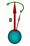
If now many nuclei of hydrogen atoms (protons) are placed in an external magnetic field, then they will begin to try to navigate along this magnetic field like the arrows of compasses. However, during such a reorientation, the nuclei will begin to precess (as the gyroscope axis precesses when trying to tilt it), because the magnetic moment of each nucleus is associated with the mechanical moment of the nucleus, with the presence of the spin mentioned above.
Suppose a hydrogen core was placed in an external magnetic field with an induction of 1 T. The precession frequency in this case will be 42.58 MHz (this is the so-called Larmor frequency for a given nucleus and for a given magnetic field induction). And if we now have an additional effect on this core with an electromagnetic wave with a frequency of 42.58 MHz, the phenomenon of nuclear magnetic resonance will occur, that is, the precession amplitude will increase, since the vector of the total magnetization of the core will become larger.
And there are one billion billions of billions of such nuclei that can precess and resonate. But since the magnetic moments of all the nuclei of hydrogen and other substances in our body interact with each other in ordinary everyday life, the total magnetic moment of the whole body is zero.
Acting on protons by radio waves, they obtain a resonant amplification of the oscillations (increase in the amplitudes of the precessions) of these protons, and upon completion of the external action, the protons tend to return to their original equilibrium states, and then they themselves emit photons of radio waves.

Thus, in an MRI device, a person’s body (or some other body or object under study) is periodically transformed into a set of radio receivers or a set of radio transmitters. Investigating in this way site after area of the body, the apparatus constructs a spatial picture of the distribution of hydrogen atoms in the body.And the higher the magnetic field strength of the tomograph - the more hydrogen atoms bonded to other atoms located nearby can be investigated (the higher the resolution of the magnetic resonance imager).
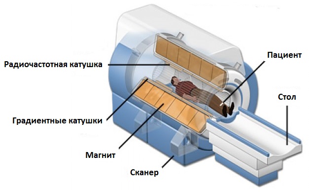
Modern medical tomographs as sources of an external magnetic field contain superconducting electromagnetscooled by liquid helium. Some open-type tomographs use permanent neodymium magnets.
The optimal magnetic field induction in an MRI machine is now 1.5 T, it allows you to get fairly high-quality images of many parts of the body. With an induction of less than 1 T, it will not be possible to make a high-quality image (of a sufficiently high resolution), for example, of the small pelvis or abdominal cavity, but such weak fields are suitable for obtaining conventional MRI images of the head and joints.

For the correct spatial orientation, in addition to a constant magnetic field, a magnetic coil also uses gradient coils, which create an additional gradient perturbation in a uniform magnetic field. As a result, the strongest resonant signal is localized more precisely in one or another section. The power and operation parameters of gradient coils - the most significant indicators in MRI - the resolution and speed of the tomograph depends on them.
See also at bgv.electricianexp.com
:
